
IVIS-XRMS Preclinical small animal imaging core facility
 Murali Anbalagan, Ph.D.
Director, Preclinical imaging core
manbalag@tulane.edu
Phone: 504-988-1563
Cell: 318-614-2171
Key Features of IVIS XRMS small animal imager
Optical and X-ray imaging
Multi-species imaging including mice and rats
High resolution, low dose digital X-ray
Exquisite sensitivity in bioluminescence
Compute Pure Spectrum (CPS) spectral unmixing for ultimate fluorescence sensitivity
Full fluorescence tunability through the NIR Spectrum
 The IVIS Lumina XRMS from PerkinElmer integrates the best in class in vivo bioluminescence and fluorescence imaging with 2D X-ray capability. The Lumina XRMS offers the flexibility to image small as well as large animals with precise optical and X-Ray overlay, giving anatomical context to the optical signal. The Lumina XRMS includes state of the art spectral unmixing for sensitive multispectral imaging to monitor multiple biological events in the same animal.
The system is equipped with up to 26 filters tunable to image fluorescent sources that emit from green to near-infrared. All Lumina Series III systems come with a novel illumination technology that effectively increases fluorescent transmission deep into the near infrared range with full transmission through 900 nm.
Standard on all IVIS instruments, absolute calibration affords consistent and reproducible results independent of magnification, filter selection from one instrument to any another IVIS instrument within an organization or around the world.
The Lumina XRMS offers the flexibility to image mice, rats and other animals up to 500-600 g in weight with an accurate optical overlay on X-ray image. The X-ray scintillator can easily be moved to effectively image mice and rats with ease. The Lumina XRMS is the only instrument that can overlay an optical signal to the X-ray image at all Fields of View. See the figures below
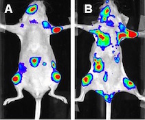
Optical overlay of bioluminescent signal with X-ray image at multiple FOV’s in mice
[3_mice]
Applications in Multimodal Imaging Precise optical and X-ray overlay brings your optical signal into anatomical context. Key applications in oncology, infectious diseases, inflammation, implant biology or any model that requires anatomical context, the Lumina XRMS Series III will offer complete and rich calibrated datasets for longitudinal studies with supporting analysis software.
Two dimensional overlaid X-ray and bioluminescent image. Red arrows highlight areas osteolysis
[bone_degradation]
IVIS Lumina Series III Software Living Image software brings IVIS technology to life by facilitating an intuitive workflow for in vivo optical, X-ray image acquisition, analysis and data organization. The software’s new design creates an intuitive, seamless workflow for researchers of all skill levels. New features include: wizard based guidance for advanced imaging protocols, spectral unmixing tools, expanded fluorescent agent database and a simplified tool palette.
[GI_Infarct]
The Small Animal Imaging Core Facility provides access to IVIS XRMS instrument, usage is restricted to authorized individuals who undergo necessary training. All users must submit the IVIS User Registration form (attached) and received Department of Comparative Medicine (DCM) approval.
Inflammation: Detection of rheumatoid arthritis with fluorescent probes (see below image)
[arthritis]
Procedure for access and usage of IVIS XR equipment
Location:
The IVIS XRMS unit is housed in the DCM facility.
Training:
Appropriate training must be obtained prior to using the IVIS. Training is provided by Dr. Murali Anbalagan (manbalag@tulane.edu).
IACUC
All users must have an approved IACUC protocol, providing experimental design details.
User Registration Form
The completed and signed User Registration Form should be submitted to Dr. Murali Anbalagan via e-mail at manbalag@tulane.edu in order to qualify for access and online sign up. Once training and registration are completed, online access for sign up and access to IVIS will be activated.
Access to the Small Animal Imaging Facility
Pending receipt of the User Registration Form and approval by DCM, your punch pad key access code for the imager will be activated for each user.
Sign up reservation
Reservations are available in blocks of one hour. Use of the instruments is available 24 hours a day/7 days a week. Reservations must be made through the calendar. Users will be added to the authorized user list upon submission of the User Registration Form. An account number is needed for reservations. If this account is invalid, the default account number on User Registration Form will be charged automatically. Please note that all sessions will be charged unless cancelled at least 12 hours in advance.
Signup URL: http://www.supersaas.com/schedule/TulaneDCM/DCM_Surgery_Rooms
Data cannot be stored on the IVIS local hard drive for longtime. After imaging all data must immediately be saved to a portable hard drive or disk.
Rate
Each IVIS XRMS session will be charged $100 for the first hour and $75 per hour after the first for time on the equipment. First 15 min is NOT charged - considered equipment set-up time.
Reserved sessions will be charged regardless of usage unless the reservation is cancelled at least 12 hours in advance. Charges will be automatically posted on the PI account provided in the User Registration form.
PI responsibilities
It is the user and PI responsibility to:
Follow all procedures
Follow carefully the IVIS SOP provided during training
Sign the user log and note any malfunction or abnormal condition of the equipment
Ensure the room and equipment are left clean and in standard configuration
Report any issue or problem immediately to Dr. Murali Anbalagan via email (manbalag@tulane.edu)
Equipment damage because of misuse or negligence will result in losing the access
Shared small animal imaging instruments pose a significant risk of cross contamination in the event of a disease outbreak within the animal facility. To minimize this risk, the users have to disinfect the instruments both before and after use. Failure to adhere to these procedures will result in retraining in the first instance, and suspension of instrument access after repeated infractions.
