
At the Tulane Advanced Imaging Center (TAIC), we understand that undergoing an MRI can feel unfamiliar or even a little intimidating. That’s why our warm, professional staff are here to guide you through every step of the process—from answering your questions to ensuring your comfort throughout the exam. Whether you're visiting for clinical research or diagnostic imaging, our goal is to provide a safe, supportive, and informative experience. Below, you'll find details to help you prepare and understand what to expect during your MRI visit.
MRI Exams
Magnetic resonance imaging (MRI) is a non-invasive, painless technology that uses a strong magnetic field and radio waves to create detailed images of organs and tissues in the body. Most MRI machines are large, cylindrical magnets that create a strong magnetic field to interact with hydrogen atoms in the body. This, combined with radiofrequency pulses, produces detailed cross-sectional images or "slices" of anatomy. MRI can also create 3D images for a full view from multiple angles. At TAIC, MRI is frequently used for clinical trials and monitoring interventions.
What will I be expected to do during MRI testing?
You will lie down on the MRI table, study specific MRI coils and accessories will be set up, and then the table will slide into the bore of the MRI machine. To ensure clear images, you’ll need to remain as still as possible throughout the scan, which can last between 30 and 90 minutes, depending on the area being imaged and the study task. Participants are often asked to hold their breath for cardiac or abdominal exams. You may be asked to perform specific tasks or respond to stimuli for fMRI exams. It is important to stay awake during the exam.
Will I need to undress to enter the MRI machine?
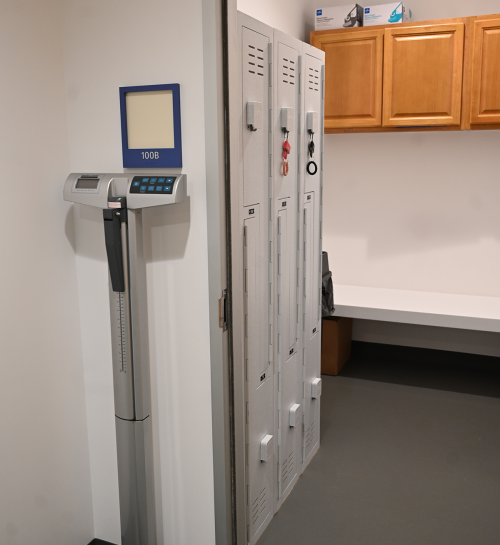
If wearing clothing with metal buttons, clasps, underwire, or zippers, you will be asked to change into a gown. Try to wear clothing without metal. You’ll also need to remove any metal items, including jewelry, glasses, underwire bras, watches, hair pins/clips, and belts before entering the MRI. Any personal belongings can be securely stored in lockers provided in the changing room.
Will I need an IV injection with contrast dye?
Some cardiac studies required an injection of MRI contrast.
You do not need to fast before your MRI. Try to only eat a small meal, if needed, before a contrast enhanced MRI.
Will I be exposed to radiation during an MRI?
MRI is a safe imaging method because it doesn’t use the ionizing radiation found in x-rays and computed tomography (CT) scans. This makes MRI ideal for cases where frequent imaging is needed, especially brain-related diagnoses and treatments.
Depending on the specific study, scans usually take from 30 to 90 minutes depending on the size of the area being scanned and the number of images being taken
What if I have a metal implant or fragments in my body?
Having metal in your body doesn’t automatically rule out an MRI scan, but it’s essential to inform the research team. They will ask about your medical history to determine if an MRI is safe for you. In some cases, an MRI may not be recommended. For instance, people with certain implants, especially those containing iron—like pacemakers, nerve stimulators, defibrillators, insulin pumps, cochlear implants, brain stimulators, or capsule endoscopy devices—should avoid MRI machines.
What if I’m a little claustrophobic?
If you experience claustrophobia, please inform the researchers. TAIC features a spacious scanner and offers two-way communication between researchers and participants, which can help ease claustrophobia for some individuals. Additionally, TAIC has a mock MRI scanner to help participants acclimate to the machine, its procedures, and the sounds it produces. To further reduce discomfort, visualization techniques are available, along with supportive options such as listening to music, watching videos, or using eye covers. A call button will be given for reassurance.
Should I consider taking medication like Valium if I'm feeling nervous?
For TAIC studies, we recommend avoiding tranquilizers and certain mood-altering medications, as they can affect brain activity and potentially influence scan results. Researchers will discuss your medications before you take part in a study.
Cardiac Exams
How should I prepare for a cardiac MRI?
You may be asked to shave your chest before a cardiac MRI to place patches for the cardiac leads. Try not to eat a large meal before your cardiac MRI exam. Try to wear clothing without metal. You may be asked to change into a gown.
Will I need IV injection with contrast dye?
Some cardiac exams at TAIC require a contrast injection. If contrast is needed, an IV will be placed into the arm. MRI contrast followed by saline solution will be administered during the cardiac MRI exam.
fMRI Exams
How is an fMRI different than an MRI?
Functional magnetic resonance imaging (fMRI) is a type of MRI that helps assess brain function by measuring and mapping brain activity. A most common fMRI method, called BOLD fMRI, works by detecting changes in blood flow and oxygen levels in the brain that happen when certain areas become more active. When a brain region is in use, it needs more oxygen, which increases blood flow to that area. By capturing these changes, fMRI shows which parts of the brain are active during specific tasks or activities.
Why use fMRI scans to reveal my brain activity?
An fMRI shows patterns of brain activity, helping researchers understand how different parts of the brain handle things like movement, language, memory and cognition. This technique has been key in identifying areas involved in thinking and movement, mapping brain connections, and studying changes in brain activity in conditions like stroke, brain injury, and diseases that affect the brain.

Will I need IV injection with contrast dye?
No contrast dyes or IV injections are used in fMRI studies.
