Tulane Cancer Center (TCC) was founded as a matrix center in 1993, a resource for supporting cancer-focused basic research and clinical faculty from home departments across the University.
Laboratories
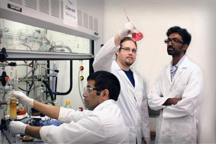
The TCC currently operates and maintains approximately 39,214 sq. ft. of active research laboratory space in the Louisiana Cancer Research Center and J. Bennett Johnston (JBJ) buildings. Research laboratories include wet bench lab bays, tissue culture, offices and shared equipment available to Cancer Center members. Also included are shared conference rooms at each site for internal lab meetings and external research collaboration meetings.
Tulane Cancer Center Shared Resources/Cores
- Common Equipment - High-end common equipment is located at both the LCRC and JBJ buildings for cancer center members to utilize for their research.
- Cell Analysis Core - The Cell Analysis Core is a customer-oriented service dedicated to supporting the research needs of the members of the Louisiana Cancer Research Consortium (LCRC). The facility is designed to collaborate with the investigators and contribute to all aspects of the research process, including consultation in experimental design, data analysis and storage, troubleshooting, interpretation of results, and the preparation and production of presentation graphics. The CACF is currently equipped with the following instrumentation:
- BD FACS Aria III - High-speed sorter that is capable of sorting up to 4 different cell populations into a variety of tubes or a single population directly into multi-well plates under sterile or non-sterile conditions based on the application.
- Cytek Aurora 4 Laser - a spectral flow cytometer equipped with four lasers (405, 488, 561, 635 nm). The Aurora’s advanced technology allows the detection of the full emission spectrum, which enables it to distinguish between overlapping dyes such as GFP and FITC or APC and CY 647.
- Next Generation Sequence Analysis Core –The NGS Core provides comprehensive standard and custom data analysis pipelines and assistance with data interpretation of a broad array of high-throughput sequencing applications, including RNA-seq, miRNA-seq, ATAC-seq, ChIP-seq, DNA-seq (Whole Genome/Exome) and CLASH-seq. Equipment includes 12 MacPro/iMacPro computers for local analyses and Tulane's High-Performance Computer for ultra-fast processing of standard and large data sets.
- Organoid Core - The long-term goal of the Tulane Cancer Center Organoid Core is to generate novel pre-clinical models of breast, lung, and pancreatic cancer as translational platforms for investigators to advance their research. The Core is located on the third floor of the J. Bennett Johnston Building (JBJ) in New Orleans, Louisiana. Currently, the Core is focused on assisting investigators who have already created their own pre-clinical models, or who require assistance using organoid models obtained through other sources to characterize their samples using the shared resources offered by the Core. As of July 1, 2025, the Core is available to assist with 2-D high-throughput screening and 3-D organoid screening by live-cell imaging (Incucyte SX5). In collaboration with Dr. Seagroves, bio-imaging of pre-clinical, immunocompromised animal models is offered using the IVIS Lumina III (Revvity) instrument.
