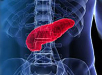
 The Hepatobiliary & Pancreas Disease Clinic provides unique diagnostic and treatment options that are available only at leading referral centers, including Endoscopic Ultrasound (EUS), Therapeutic Endoscopic Retrograde Cholangiopancreatography (ERCP) and advanced surgical techniques such as the Whipple procedure. Additionally, we offer unique services such as pancreas pain management, and specialized surgical services for the repair of bile duct injuries, pancreas preservation surgery, and pancreas transplantation.
Our diagnostic and treatment options include:
- Abdominal CT
- Abdominal MRI
- Abdominal Ultrasound
- Balloon Dilation
- Central Pancreatectomy
- Cholecystectomy
- Cholangiography
- Distal Pancreatectomy
- Endoscopic Retrograde Cholangiopancreatography (ERCP)
- Endoscopic Retrograde Sphincterotomy (ERS)
- Endoscopic Ultrasound (EUS)
- Enucleation
- Fine Needle Aspiration
- Hepatobiliary Iminodiacetic Acid (HIDA) Scan
- Laparoscopic (minimally invasive) Surgery
- Robotic Surgery
- MRCP (magnetic resonance cholangiopancreatography)
- Pancreas Transplant
- Pancreaticoduodenectomy
(Whipple procedure) - Pancreatic Resection
- Percutaneous Therapy for Bile Stricture
- Percutaneous Transhepatic Cholangiogram (PTC)
- Peustow Procedure
- Stent Placement
- Whipple Procedure
Abdominal CT - Computerized Tomography (CT) is an imaging modality that generates detailed cross-sectional images of the pancreas, liver, gallbladder and other organs. Specialized pancreas protocol CT scans improve pancreas images compared to the standard CT scans.
Abdominal MRI- Magnetic Resonance Imaging (MRI) is an imaging modality that produces precisely detailed images of the pancreas, bile ducts, liver and gallbladder by using magnetic field and radio waves to image the organs.
Abdominal Ultrasound – ultrasound utilizes sound waves to create an image of the pancreas, liver, gallbladder and other organs. If gallstones are present they will be visible on the ultrasound. Intraoperative ultrasound is used during surgery to help surgeons locate areas of cancerous tissue within the biliary tract (gallbladder and bile ducts that store and carry bile).
Balloon Dilation - Therapeutic Endoscopic Retrograde Cholangiopancreatography (ERCP) with balloon dilation is often used as a method of treating bile duct strictures. If the stricture completely blocks the bile duct a balloon on the tip of a catheter is passed through the ERCP endoscope and is inflated at the site of the stricture, enlarging the blocked duct. After dilation a stent is passed through the ERCP endoscope and placed at the point of stricture to keep it open.
Central Pancreatectomy- This procedure is performed if there is a benign tumor in the neck of the pancreas. The focus of the surgery is to remove any tumors in the area while preserving as much of the pancreas as possible, as well as avoiding complications such as diabetes and malabsorption of nutrients.
Cholecystectomy – surgical removal of the gallbladder is referred to as a cholecystectomy. This is the most common treatment methodology for gallstones. The procedure can be performed using a laparoscopic (minimally invasive) procedure, robotic, or traditional, open procedure.
Cholangiography– A cholangiography is an x-ray imaging modality that uses contrast dye to highlight and view any obstructions or narrowing of the bile ducts. into the bile ducts by advancing a needle through the skin and liver in order to inject contrast dye into the bile ducts, or by endoscopically placing a catheter where the bile duct enters the small intestine, followed by an x-ray. The contrast dye can be introduced by one of several ways:
- Percutaneous Transhepatic cholangiography (PTC) – the contrast dye is injected through the skin into the bile ducts within the liver using ultrasound to guide the needle. X-rays are taken to identify any narrowing or blockages in the biliary system.
- Intraoperative Cholangiography – the contrast dye is injected into the bile duct during gallbladder surgery. X-rays are taken to identify any narrowing or blockages in the biliary system.
- Endoscopic Retrograde Cholangiopancreatography (ERCP) – the contrast dye is injected into the common bile duct and pancreatic duct through a catheter that is passed through an endoscope. X-rays are taken to identify any narrowing or blockages in the biliary system
- Magnetic resonance cholangiopancreatography (MRCP) - MRCP uses magnetic resonance imaging (MRI) instead of x-rays to produce an image of the bile ducts, pancreatic duct, and gallbladder. No contrast dye is used.
Distal Pancreatectomy – A Distal Pancreatectomy is a pancreas surgery that is used to remove tumors located in the body and the tail of pancreas, leaving the head of the pancreas intact. Unless the tumor is identified as a low-grade malignancy or benign disorder of the tail of the pancreas the spleen is generally removed during this surgery as well due to its close association with the tail of the pancreas.
Endoscopic Retrograde Cholangiopancreatography (ERCP) - ERCP is an advanced endoscopic technique that combines endoscopy and fluoroscopy to diagnose and treat problems associated with the pancreatic duct system and biliary system. This procedure is performed by a gastroenterologist with specialized training in ERCP. Using an endoscope (a flexible, fiber-optic scope), the gastroenterologist injects contrast dye into the pancreatic duct or bile ducts in order to highlight the ducts and assist in taking x-rays. Endoscopic Sphincterotomy (ERS) can be performed as part of a ERCP procedure, during which instruments are inserted through the endoscope in order to enable the removal of stones, place stents, or expand narrowed biliary or pancreatic ducts.
- Gallstone Removal - used to remove pancreatic or bile duct stones. A sphincterotomy is sometimes performed at the same time.
- Stent placement – allows for the placement of a device to keep the pancreatic duct or bile duct open.
- Balloon dilatation – allows a narrowed pancreatic duct or bile duct to be opened.
Endoscopic Ultrasound (EUS) – EUS is an advanced diagnostic imaging option that is used to visualize the structures of the pancreas and biliary system. Traditional ultrasound imaging process produces views of specific organs by sending sound waves back and forth through a transducer that is placed on the skin. Endoscopic Ultrasound combines endoscopy (a flexible, fiber-optic scope) and ultrasound in order to produce higher resolution images and information about specific organs by placing a small ultrasound transducer on the tip of the endoscope. EUS provides greater detail than traditional ultrasound because of the proximity of the ultrasound device to the pancreas or bile ducts. EUS can also obtain detailed information about the layers of the intestinal wall and the corresponding lymph nodes and blood vessels. EUS is often used in conjunction with Endoscopic Retrograde Cholangiopancreatography (ERCP).
Endoscopic Retrograde Sphincterotomy (ERS) – Performed during an ERCP procedure, ERS inserts instruments through the endoscope in order to enable the removal of stones, place stents, or expand narrowed biliary or pancreatic ducts.
- Gallstone Removal – used to remove pancreatic or bile duct stones. A sphincterotomy is sometimes performed at the same time.
- Stent Placement – allows for the placement of a device to keep the pancreatic duct or bile duct open.
- Balloon dilatation – enlarges and opens a narrowed pancreatic duct or bile duct.
Enucleation - Many functional pancreatic islet tumors are small and are located on the surface of the pancreas contained by a lining around them that separates them from the pancreas. Enucleation surgery allows for the removal of these tumors without removing any pancreatic tissue.
Fine-Needle Aspiration (FNA) – Fine Needle Aspiration is a biopsy procedure that guides a small needle through the skin and abdomen to the appropriate location so that a small sample can be removed and examined. Ultrasound or a CT scan is used to direct the needle.
Hepatobiliary Iminodiacetic Acid (HIDA) Scan – A hepatobiliary iminodiacetic acid scan is an imaging procedure that tracks the production and flow of bile from the liver to the small intestine. Radioactive contrast material is injected and then allowed to circulate to the liver where it is excreted into the biliary system and stored by the gallbladder.
Laparoscopic and Robotic Surgery - Laparoscopic surgery is a minimally-invasive surgical technique through which several small incisions in the abdomen allow the insertion of surgical instruments and a video camera. The camera illuminates the surgical field and sends an image to a video monitor, providing the surgeon an enhanced view of the surgical site. The surgeon uses the monitor image to perform surgery by manipulating the surgical instruments through the operating ports. Laparoscopic and robitic surgery may result in fewer complications and improved recovery time as compared to the traditional (open) surgical approach.
Magnetic Resonance Cholangiopancreatography (MRCP) - MRCP is a magnetic resonance imaging (MRI) exam that produces detailed images of the hepatobiliary and pancreatic systems, including the liver, gallbladder, bile ducts, pancreas and pancreatic duct without the use of contrast dye. MRCP produces images similar to an ERCP and may be used in place of ERCP for diagnostic purposes if therapeutic interventions such as stent placement are not required.
Pancreas Transplant – As part of our multidisciplinary approach to the treatment of diseases and disorders of the pancreas and biliary tract, our team works closely with the surgeons and staff of the Tulane Transplant Institute.
Pancreaticoduodenectomy – Also known as the Whipple procedure, the pancreaticoduodenectomy is the most common procedure for pancreatic tumors. During the procedure the surgeon removes of the head of the pancreas, most of the duodenum (the first part of the small intestine), a portion of the bile duct, the gallbladder, and associated lymph nodes. In some cases the entire duodenum is removed as well as a portion of the stomach.
Pancreatic Resection - A pancreatic resection is a surgery to remove a tumor from the pancreas and is performed on patients in which the tumor is localized and meets specific criteria for the stage and classification of the malignancy. The surgical focus of pancreatic resections performed at the Pancreas and Biliary Center is to preserve as much pancreas tissue as possible.
Percutaneous Therapy for Bile Stricture - If Therapeutic Endoscopic Retrograde Cholangiopancreatography (ERCP) is not indicated for placing a stent to open a biliary stricture another option is to use a percutaneous (through the skin) procedure to dilate the stricture and enable stent placement.
Percutaneous Transhepatic Cholangiogram (PTC) – Percutaneous Transhepatic Cholangiogram is an x-ray image of the bile duct system. During the exam a thin needle is inserted through the skin (percutaneous) and through the liver (transhepatic) into a bile duct. A contrast dye is injected and the bile duct system is outlined on x-rays. The procedure is generally reserved for patients who have undergone an unsuccessful ERCP procedure.
Puestow Procedure – The Puestow procedure (lateral pancreaticojejunostomy) is a surgical technique used in the treatment for pain associated with chronic pancreatitis. During the procedure the pancreas is exposed and the main pancreatic duct is opened from the head to the tail of the pancreas. The opened pancreatic duct is then connected to a loop of small intestine so that the pancreas drains directly into the intestines. Our pancreas surgeons employ many advanced techniques such as the Puestow and Frey procedures, always individualizing treatment to individual circumstances.
Stent - A stent is a thin, tube-like structure that is used to open and support a narrowed portion of the pancreatic duct or bile duct, and prevent the reformation of the stricture. Stents may be made of plastic or metal. Biliary system stent placement can be performed during endoscopic retrograde cholangiopancreatography (ERCP).
Whipple Procedure – The Whipple procedure, also known as pancreaticoduodenectomy, is the most common procedure for pancreatic tumors. During the procedure the surgeon removes of the head of the pancreas, most of the duodenum (the first part of the small intestine), a portion of the bile duct, the gallbladder, and associated lymph nodes. In some cases the entire duodenum is removed as well as a portion of the stomach.
