Interventional Pulmonology is a branch of medicine that uses minimally invasive procedures aimed at diagnosing and treating various diseases, including lung cancer, pleural diseases, and many types of complex airway and lung disorders.
Interventional Pulmonology: Why Choose Tulane?
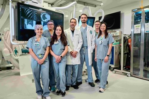
The Interventional Pulmonology team at Tulane uses the latest technology to provide patients with alternatives to more invasive surgical procedures. Our Interventional Pulmonologists work alongside your primary care physician, your pulmonologist, oncologists, thoracic surgeons, radiation oncologists, radiologists, otolaryngologists, and anesthesiologists to provide comprehensive care to a complex patient population.
Tulane Interventional Pulmonology has a dedicated procedure suite equipped with the most advanced technologies, including rigid and flexible bronchoscopy, cryobiopsy, navigational bronchoscopy, pleuroscopy, a cone beam CT scanner, and on-site cytopathology support. Our team consists of the interventional pulmonologist, a group of dedicated anesthesiologists, and experienced nursing and respiratory staff focused on patient care.
Conditions We Treat
- Airway obstructions, including foreign bodies
- Central airway collapse (tracheobronchomalacia)
- Emphysema
- Interstitial lung disease
- Lung nodules/masses
- Mediastinal adenopathy or masses
- Pleural disease
- Pneumothorax
Tests, Treatments and Services We Offer
- Flexible Bronchoscopy
- Rigid Bronchoscopy
- Airway tumor/tissue ablation
- Endobronchial Ultrasound (EBUS)
- Electromagnetic Navigational Bronchoscopy
- Endobronchial Valve Placement
- Cryobiopsy
- Balloon Dilation
- Airway Stents
- Thoracentesis
- Tunneled Pleural Catheter Placement
- Pleuroscopy (Medical Thoracoscopy)
- Percutaneous Dilation Tracheostomy
Minimally Invasive Procedures
Minimally invasive techniques are aimed at decreasing blood loss, reducing pain, as well as minimizing time to recovery when compared to traditional surgical techniques. Patients often spend less time in the hospital and are able to return to their daily activities sooner.
Procedures
What is Flexible or Rigid Bronchoscopy?
Flexible and rigid bronchoscopy is performed by inserting a small flexible or rigid scope with a camera attached into the airways. This procedure can diagnose and treat a wide array of diseases including airway blockages, infections, and lung cancer. The patient is normally asleep during this procedure and is sedated by our anesthesiologists.
What is Endobronchial Ultrasound (EBUS)?
Endobronchial ultrasound is a minimally invasive procedure using an EBUS bronchoscope, which is a flexible bronchoscope combined with an ultrasound. Using this bronchoscope, we can examine and biopsy suspicious lymph nodes adjacent to the airways. Using this technique, we can diagnose lung cancer or many inflammatory and infectious diseases without performing surgery. Our patients normally go home the same day.
What is Electromagnetic Navigational Bronchoscopy?
Electromagnetic Navigational Bronchoscopy (ENB) is a minimally invasive procedure allowing the bronchoscopist to use GPS-like technology to create a 3-D map of your airways, allowing them to navigate to your lung nodule and obtain tissue without surgery. This technology can biopsy lung nodules that cannot be obtained through traditional bronchoscopy, place fiducial markers, and help diagnose early stage lung cancer, allowing for rapid treatment. Patients normally go home the same day.
What is an Endobronchial Valve?
An Endobronchial valve (EBV) is a small medical device that can be implanted in an airway to treat airway leaks and to help certain patients with emphysema breath more easily. In patients with severe emphysema, EBV placement usually requires a 3 day hospital stay for monitoring.
What is Cryobiopsy?
Cryobiopsy is another way to obtain tissue from the lungs. It uses a cryoprobe, or a freezing probe, to obtain tissue while protecting the architecture of the airways, allowing pathologists to more accurately diagnose diseases of the lung, including Interstitial lung disease (ILD). Patients normally go home the same day.
What is Balloon Dilation?
Using the bronchoscope, a small balloon can be inflated, improving the airway narrowing caused by tumors, inflammatory conditions, or other airway diseases. This can provide immediate improvement in airway narrowing. If narrowing continues, balloon dilation can pave the way for stent placement, if necessary.
What are airway stents?
Tracheobronchial airway stents are small tubes that are placed into the airway to keep the airways open. They can be made of silicone or metal and can be permanent or temporary.
What is a Thoracentesis?
The physician uses an ultrasound to locate a pocket of fluid. Next, a small needle with a catheter (much like an IV) is inserted into the chest cavity (the area between the lungs and the chest wall) to obtain suspicious pleural fluid. Once obtained, the fluid is sent for analysis and can help diagnose cancer, infection, or many inflammatory processes. Removing this fluid can also make the patient breathe more easily. Patients normally go home the same day.
What is a Tunneled Pleural Catheter?
After thoracentesis, if the pleural fluid reaccumulates and symptoms recur, we can place a more permanent catheter allowing drainage at home, either by the patient themselves or a visiting nurse, decreasing hospitalizations and improving patient comfort and quality of life. This procedure is similar to a thoracentesis and the patient normally goes home the same day.
What is Pleuroscopy (Thoracoscopy)?
Thoracoscopy is a minimally invasive procedure used to examine the chest cavity for abnormalities. A small incision is made in the chest wall and a small tube with a camera at the end is inserted. The physician can take biopsies of abnormal looking tissue and take fluid samples to diagnose a wide array of diseases.
What is Percutaneous Dilational Tracheostomy?
Percutaneous Dilational Tracheostomy (PDT) is a minimally invasive procedure that inserts a small tube into the anterior neck, normally in a loved one who is critically ill. It is usually done at the bedside in the ICU.
Our Team
Ramsy Abdelghani, MD - Interventional Pulmonologist
David Becnel, MD – Interventional Pulmonologist
Diana Espinoza Barrera, MD - Interventional Pulmonologist
Nikkie Nguyen, RN - Registered Nurse, Interventional Pulmonary Medicine
Social Media
![]() Follow Dr. Abdelghani on Twitter
Follow Dr. Abdelghani on Twitter
OR
Point your phone's camera at this QR code