
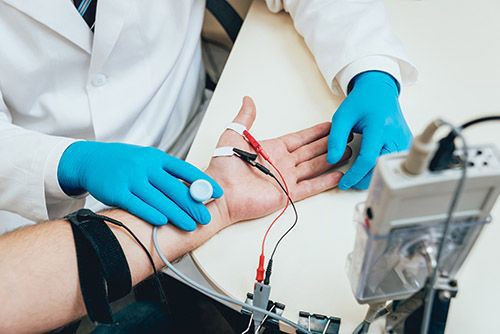
Tulane Neurology has a number of faculty members with expertise in neuromuscular disease. EMG’s are performed by either Dr. Kristina Lafaye, or Dr. Morteza Shamsnia who all have advanced training and expertise in this subspecialty of Neurology.
Neuromuscular disease is very common and can include peripheral neuropathy as well as muscle dysfunction known as myopathy. There are a large number of patients with numbness and pain related to peripheral neuropathy with diabetes mellitus the most common cause of such a presentation. There is usually sensory loss with numbness and tingling as the primary manifestation. However, burning or sharp and shooting so-called neuropathic pain can make neuropathy quite uncomfortable for a significant percentage of patients with neuropathy. Neuropathy typically affects the distal extremities, i.e. the feet and hands, more than the proximal regions such as the hips and shoulders. This has been termed a “stocking-glove” pattern to the neuropathy as opposed to the most common forms of myopathy where proximal involvement tends to be greater than distal involvement. Some neuropathies primarily affect motor function with weakness and some are combined sensorimotor neuropathies.Read More
There are a number of potential causes for neuropathy outside of diabetes and up to one half of patients have no readily identified cause. Neuropathy can be seen secondary to deficiency conditions such as vitamin B12 deficiency and hypothyroidism, as a complications of infection such as HIV and syphilis as well as Lyme disease, in inflammatory conditions such as systemic lupus erthythematosus and polyarteritis nodosa, and as what is termed as a remote effect of malignancy. There are also medications which can promote neuropathy such chemotherapeutic agents for cancer. In addition, there are familial forms of neuropathy. There can be an acute neuropathy associated with significant weakness in an ascending fashion termed Guillain-Barre syndrome. This is a neurological emergency as there can be life-threatening respiratory insufficiency associated with this “ascending paralysis” along with cardiovascular instability.
The evaluation includes a careful history and exam with the determination of neuropathy often made clinically. Additional testing may include electromyography with nerve conduction velocity measurements to determine the general type of neuropathy, either demyelinative, axonal, or combined. There are routine blood studies that are performed as part of the investigation of neuropathy including a vitamin B12 level , thyroid profile as well as assessment for diabetes or glucose intolerance. Evaluation for occult malignancy might also be in order as well as evaluation for a possible connective tissue disorder. For certain patients, a nerve biopsy may be indicated to determine the cause with special staining of the specimen.
Treatment of neuropathy depends on the particular condition associated with the neuropathy. Efforts need to be made to promote functional independence and to protect against falls as some patients develop gait instability related to what is termed a “sensory ataxia”. Diabetic patients with neuropathy tend to benefit from improved blood sugar control. Those with deficiency conditions such as vitamin B12 deficiency or thyroid hormone deficiency will benefit from replacement therapy. Effective treatment of the neoplastic process might help those patients with a neuropathy associated with their cancer. Guillain-Barre syndrome responds to acute intervention with either intravenous gammaglobulin therapy or to plasmapheresis.
Myopathies include familial forms such as muscular dystrophy. There can be muscle weakness and muscle pain related to certain medications such as statin agents used commonly to lower the cholesterol level. Some patients have inflammatory conditions such as polymyositis and dermatomyositis. Certain infections can cause myopathy and it can be associated with HIV related disease. The evaluation includes electromyography (EMG) to look for typical myopathic findings. Many patients will require a muscle biopsy to better characterize the cause of the myopathy and to determine the optimal therapeutic approach. This might include steroid therapy for inflammatory myopathies such as polymyositis.
Neuromuscular junction disorders include myasthenia gravis and Eaton-Lambert syndrome. Myasthenia gravis is an autoimmune disorder of the junction between the innervating nerve and the particular muscle. It is autoimmune in nature with antibodies to the acetylecholine (neurotransmitter) receptor blocking the innervations of the muscle resulting in weakness of the affected muscles. Eye muscles are often involved with drooping of the eyelids (ptosis) as well as difficulty looking in a particular direction resulting in double vision. There can be bulbar musculature involvement with slurring of speech difficulty swallowing, and respiratory muscles can be involved which could lead to intubation and respiratory support during an acute attack. There can also be pronounced generalized weakness in certain patients. The diagnosis is clinical with electrophysiological support showing decremental innervations of a particular muscle with repetitive stimulation. There can be antibodies to acetylcholine antibodies, available commercially, and this is positive in up to 90% of patients and can help to support the diagnosis. Empiric treatment of this presumptive disorder with a cholinesterase inhibitor, such as (edrophonium) Tensilon, can result in a dramatic objective response in certain patients that helps to strongly support the diagnosis. However, the Tensilon test has become less popular because of potential cardiac side effects in certain susceptible individuals.
Most patients with myasthenia gravis have some form of abnormality of their thymus gland within their chest termed thymic hyperplasia and this is seen in up to three-quarters of patients by CT imaging of the chest. A smaller percentage, roughly 10%, have an actual thymus gland tumor called a thymoma. Removal of the thymus gland can be a therapeutic option for patients especially if there is a thymic gland tumor or they are having progression of their disease despite medical therapy. Standard therapy has been with oral pyridostigmine (Mestinon) which increases the level of acetylcholine at the neuromuscular junction by inhibiting the acetylcholine breakdown enzyme cholinesterase. This can be quite effective for certain individuals with myasthenia gravis who have a relatively benign course of the disease. For those who do not respond adequately to a cholinesterase inhibitor, then steroid therapy may be indicated with the caveat that initiation of such therapy can sometimes make the disease worse rather than better at the time of initiation. Other immunosuppressive therapy, such as azothioprine, methotrexate, cyclophosphamide or mycophenolate mofetil (Cellcept) may be indicated in progressive cases.
There can be acute deterioration in myasthenia gravis termed myasthenic crisis. This can be life threatening and calls for careful attention to the respiratory function with respiratory support as indicated and a determination that the worsening is not related to overmedication of the patient. In such inistances, plasmapheresis or intravenous gammaglobulin can be quite beneficial in promoting remission of the decompensated condition. Such patients are generally managed in the intensive care setting and careful monitoring of pulmonary function is indicated for any patient that appears to be susceptible to myasthenic crisis.
Lambert-Eaton syndrome, also known as myasthenic syndrome, is a neuromuscular junction disorder in which 85% of patients have antibodies to voltage-gated calcium channels. These antibodies against presynaptic transmission, at the neuromuscular junction, can lead to generalized weakness with both the arms and legs often prominently involved. Typically, there are hypoactive or absent deep tendon reflexes and dry mouth. At least 60% of patients have an associated malignancy with small cell lung carcinoma most frequently identified. Thus, there are often both autoimmune as well as paraneoplastic aspects to this disease. The ocular muscles are not usually involved and this tends to make this illness readily distinguishable from myasthenia gravis. Furthermore, unlike myasthenia gravis, where there is a decremental response to repetitive nerve stimulation by compound muscle action potential on EMG, Lambert-Eaton syndrome is characterized by an incremental response. This helps to support the diagnosis when clinically suspected and there can also be support from detection of the antibodies to the voltage-gated calcium channels. The major therapeutic approach, if identifiable and feasible, is to remove the associated malignancy. Other than this, the agent 3,4-aminopyridine, a potassium channel blocker, can provide some benefit for the weakness and plasma exchange with removal of the antibodies may also be of some potential benefit.
Motor neuron disease can encompass a spectrum of presentations including spinomuscular atrophy (lower motor neuron presentation characterized by weakness, atrophy and fasciculations of the involved muscles), primary lateral sclerosis (upper motor neuron presentation characterized by spastic weakness with hyperreflexia and positive Babinski reflex), progressive bulbar atrophy with bulbar type dysarthia and swallowing difficulty and potential for respiratory compromise), or the combination of these sub-types, which is most common and is known as amyotrophic lateral sclerosis (ALS). This has been known among the lay public as Lou Gehrig’s disease and is typically associated with a progressive deteriorating course of illness with diffuse severe weakness resulting in the victim being wheelchair bound with the increasing need for respirator and nutritional support. It is usually a sporadic illness but can be familial. In the diagnostic assessment, there is often MR imaging of the cervical spine as well as the brain in light of the serious prognosis implications of such a diagnosis. Atrophy and fasciculations of the tongue place it above the cervical spine in terms of involvement as sometimes a combination of compressive cervical radiculopathy and myelopathy can mimic ALS. EMG is usually performed to help confirm the diagnoss and there is typically a denervation/reinervation pattern in the involved musculature. Such findings in the tongue is particularly troublesome in terms of prognosis as the bulbar involvement is a harbinger of progressive speech, swallowing and respiratory compromise. Treatment is mainly supportive to protect against the progressive immobility associated with the disease along with respiratory and nutritional support that can include being placed on a ventilator and having a percutaneous endoscopic gastrostomy (PEG) tube placed for nutritional support. The agent Rilutek is released by the FDA for this disorder, but the response tends to be quite limited.
